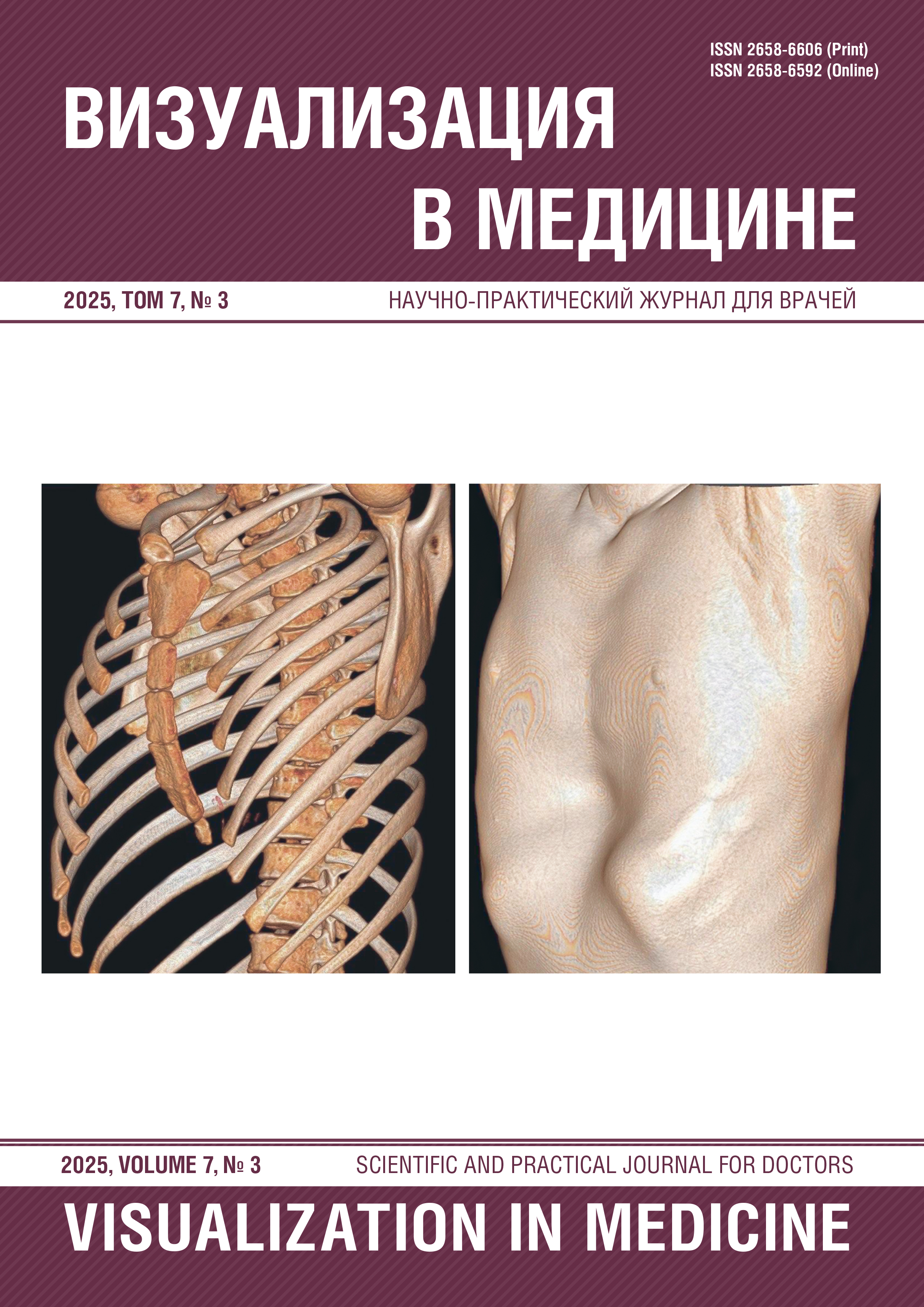
Radiation diagnostics of funnel-shaped deformation of the chest in children
Abstract
The most common congenital anomaly of the anterior chest wall is a funnel-shaped chest deformity. A funnel-shaped chest
deformity is not only an aesthetic problem, but also a medical and social problem that requires an interdisciplinary approach. In severe
cases, the deformity can lead to respiratory and heart problems, gastrointestinal issues, and secondary complications such as kyphosis
or scoliosis, as well as serious psychological problems related to quality of life. The role of radiation diagnostics in this case is key and
indispensable at all stages — from initial examination to postoperative control, as it is a complex of methods, including radiography,
computed tomography and magnetic resonance imaging, which allow not only to visualize the defect, but also to quantify its severity by
calculating the Haller index,the index of correction and angle of inclination of the sternum allows you to objectively assess the degree
of compression effect on the mediastinal organs, heart, main vessels and pulmonary parenchyma, identify concomitant pathology,
accurately draw up an individual plan for the upcoming surgical correction, choose the optimal size and simulate the implant, and then
conduct full-fledged postoperative monitoring to assess the correct position of the installed plate, bone consolidation, elimination of
compression of internal organs and early detection of possible complications such as structural migration
References
1. Fokin A.A., Steuerwald N.M., Ahrens W.A. et al. Anatomical, histologic, and genetic characteristics of congenital chest wall deformities. Semin Thorac Cardiovasc Surg. 2009;21(1):44–57. https://doi.org/10.1053/j.semtcvs.2009.03.001.
2. Westphal F.L., Lima L.C., Lima Neto J.C. et al. Prevalence of pertussis carinatum and pertussis excavatum in students in the city of Manaus. Brazil. J Bras Pneumol. 2009;35(3):221–226. https://doi.org/10.1590/s1806-37132009000300005.
3. Kelly RE Jr. Pectus excavatum: historical background, clinical picture, preoperative evaluation and criteria for operation. Semin Pediatr Surg. 2008;17(3):181–193. https://doi.org/10.1053/j.sempedsurg.2008.03.002.
4. Coln E., Carrasco J., Coln D. Demonstrating relief of cardiac compression with the Nuss minimally invasive repair for pectus excavatum. J Pediatr Surg. 2006;41(4):683–686; discussion 683-6. https://doi.org/10.1053/10.1016/j.jpedsurg.2005.12.009.
5. Crossland D.S., Auldist A.W., Davis A.M. Malignant pectus excavatum. Heart. 2006;92(10):1511. https://doi.org/10.1136/hrt.2005.087098.
6. Sang-Hyun S., Ung-Kyu C. Clinical significance of radiological stability in reconstructed thoracic and lumbar spine following body resection. J Korean Neurosurg Soc. 2014;56(4):323–329. https://doi.org/10.3340/jkns.2014.56.4.323.
7. Scalise P.N., Demehri F.R. The management of pectus excavatum in pediatric patients: a narrative review. Transl Pediatr. 2023;12(2):208–220. https://doi.org/10.21037/tp-22-361.
8. МР 2.6.1.0066-12. 2.6.1. Ионизирующее излучение, радиационная безопасность. Применение референтных диагностических уровней для оптимизации радиационной защиты пациента в рентгенологических исследованиях общего назначения: Методические рекомендации (утв. Роспотребнадзором 23.07.2012). M.; 2012.
9. Calloway E.H., Chhotani A.N., Lee Y.Z. et al. Three-dimensional computed tomography for evaluation and management of children with complex chest wall anomalies: useful information or just pretty pictures? J Pediatr Surg. 2011;46(4):640–647. https://doi.org/10.1016/j.jpedsurg.2010.10.013.
10. Robbins L.P. Pectus excavatum. Radiology case reports. 2015;6(1):460. https://doi.org/10.2484/rcr.v6i1.460.
11. Cartoski M., Nuss D., Goretsky M. et al. Classification of the Dysmorphology of Pectus Excavatum. J Pediatr Surg. 2006;41(9):1573–1581. https://doi.org/10.1016/j.jpedsurg.2006.05.055.