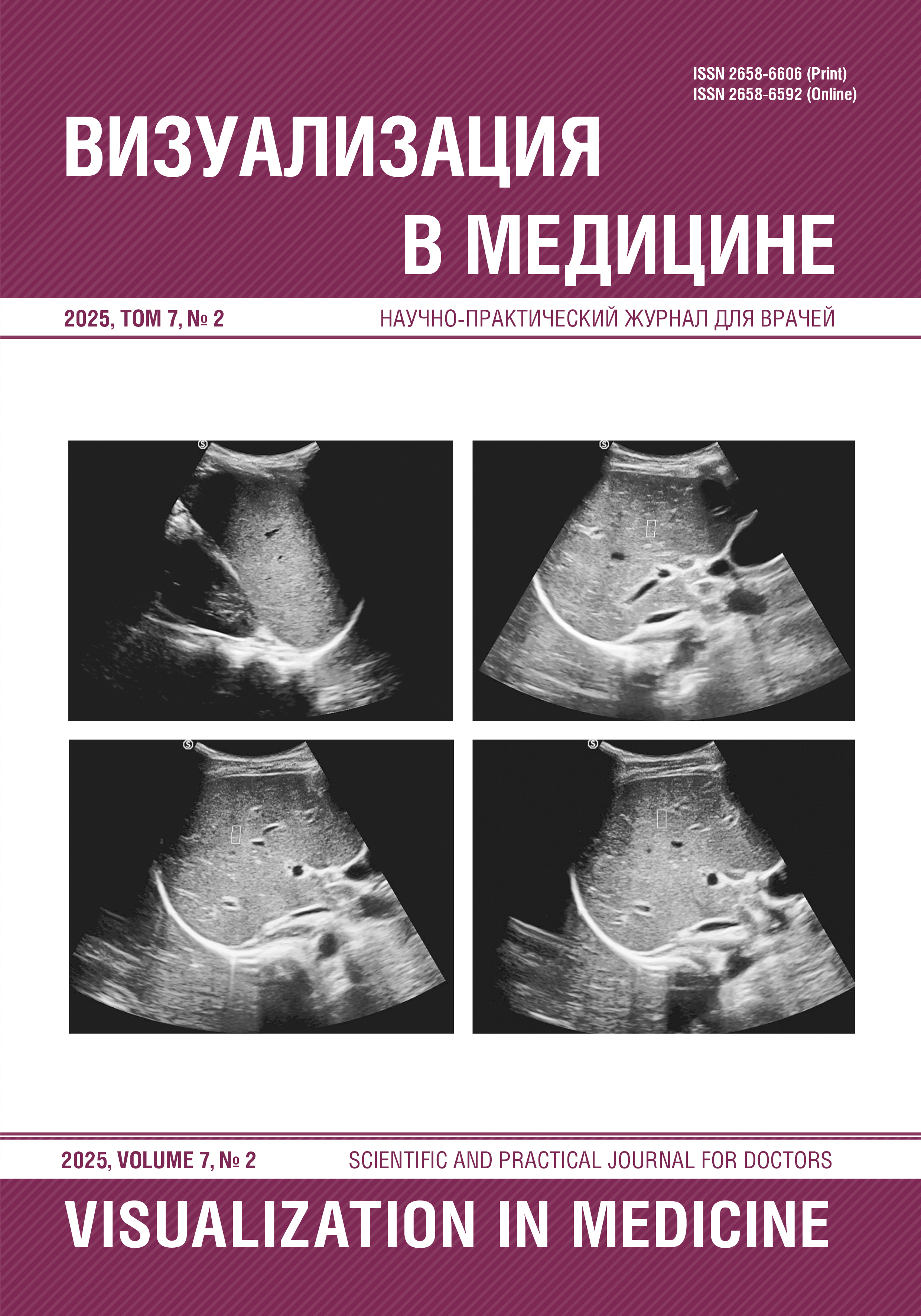
Ultrasound diagnostics of liver fibrosis in children using point shear wave elastography
Abstract
The study is devoted to the assessment of the diagnostic accuracy of the point shear wave elastography (pSWE) method in determining the stages of liver fibrosis in children. The retrospective study included 15 patients (7 boys and 8 girls) aged from 6 months to 17 years with various chronic liver diseases. All children underwent pSWE on the SIUI Apogee 6500 ultrasound device and laparoscopic liver biopsy, after which the fibrosis stage was assessed according to the METAVIR scale. Statistical analysis showed a strong positive correlation between the shear wave velocity according to pSWE and the degree of fibrosis (r = 0.850, p = 0.0001). ROC analysis demonstrated high accuracy of the method: AUC was 0.96 for F≥2, 0.94 for F≥F2-F3, 0.86 for F≥F3-F4 and 0.86 for F=F4. However, in 33.3% of cases, discrepancies were found between the pSWE results and histology. False-positive results were observed in patients with high hepatitis activity, and false-negative results were observed in patients with Crohn’s disease. Thus, pSWE demonstrates high diagnostic accuracy and can be used as a screening method for assessing liver fibrosis in children, but data interpretation requires taking into account the clinical and laboratory context.
References
Малеев В.В., Ситников И.Г., Бохонов М.С. Вопросы гепатологии: учебное пособие. В.В. Малеев, ред. СПб.: СпецЛит; 2016.
Ивлева С.А., Дворяковский И.В., Смирнов И.Ч. Современные неинвазивные методы диагностики фиброза печени у детей. Российский педиатрический журнал. 2017;20(5):300–306.
Ивашкин В.Т., Маевская М.В., Жаркова М.С., Жигалова С.Б., Киценко Е.А., Манукьян Г.В., Трухманов А.С., Маев И.В., Тихонов И.Н., Деева Т.А. Клинические рекомендации Российского общества по изучению печени и Российской гастроэнтерологической ассоциации по диагностике и лечению фиброза и цирроза печени и их осложнений. Российский журнал гастроэнтерологии, гепатологии, колопроктологии. 2021;31(6):56–102.
Соловьева Ю.А. Неинвазивные методы диагностики фиброза печени. Вестник Северо-Восточного федерального университета им. М.К. Аммосова. Серия: Медицинские науки. 2020;4(21):45–54. DOI: 10.25587/SVFU.2020.21.4.005.
European Association for the Study of the Liver. EASL Clinical Practice Guidelines on non-invasive tests for evaluation of liver disease severity and prognosis — 2021 update. J Hepatol. 2021;75(3):659-689.
Ferraioli G., Wong V.W., Castera L. et al. Liver ultrasound elastography: an update to the World Federation for Ultrasound in Medicine and Biology Guidelines and Recommendations. Ultrasound Med Biol. 2018;44(12):2419–2440.
Regev A., Berho M., Jeffers L.J. et al. Sampling error and intraobserver variation in liver biopsy in patients with chronic HCV infection. Am J Gastroenterol. 2002;97(10):2614–2618.
Bota S., Herkner H., Sporea I. et al. Meta-analysis: ARFI elastography versus transient elastography for the evaluation of liver fibrosis. Liver Int. 2013;33(8):1138–1147. DOI: 10.1111/liv.12240.
Saglam F., Beyazit Y., Sisman G. et al. Assessment of liver stiffness in patients with inflammatory bowel disease using acoustic radiation force impulse imaging. Med Ultrason. 2015;17(4):456–462. DOI: 10.11152/mu.2013.2066.174.ass.
Millonig G., Reimann F.M., Friedrich S. et al. Extrahepatic cholestasis increases liver stiffness (FibroScan) irrespective of fibrosis. Hepatology. 2008;48(5):1718–1723. DOI: 10.1002/hep.22577.
Hudson J.M., Milot L., Parry C., Williams R., Burns P.N. Inter- and intra-operator reliability and repeatability of shear wave elastography in the liver: a study in healthy volunteers. Ultrasound Med Biol. 2013;39(6):950–955.
Arena U., Vizzutti F., Corti G. et al. Acute viral hepatitis increases liver stiffness values measured by transient elastography. Hepatology. 2008;47(2):380–384. DOI: 10.1002/hep.22007.
Trout A.T., Xanthakos S.A., Bennett P.S. et al. Liver shear wave speed and other quantitative ultrasound measures of liver parenchyma: prospective evaluation in healthy children and adults. Am J Roentgenol. 2020;214(3):557–565.
Barr R.G., Ferraioli G., Palmeri M.L. et al. Elastography assessment of liver fibrosis: society of radiologists in ultrasound consensus conference statement. Radiology. 2015;276(3):845–861.
Dezsofi A., Baumann U., Dhawan A. et al. Liver biopsy in children: position paper of the ESPGHAN Hepatology Committee. J Pediatr Gastroenterol Nutr. 2015;60(3):408–420.